
What Is Protein Interface Design? From Molecular Interface Recognition to AI-Driven Engineering
Published on October 28, 2026
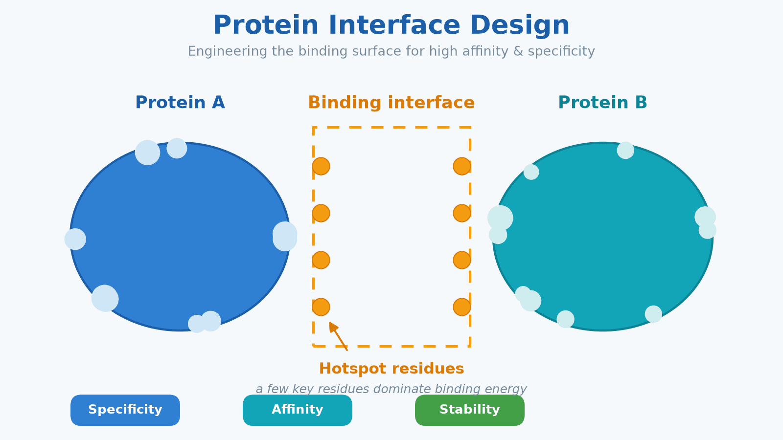
Figure 1 Protein interface: complementary binding and hotspot residues
Category: C (Directed Evolution / Protein Engineering), cross-cutting to E (Structure / Mechanism / Sequence Analysis)
Introduction
Protein Interface Design refers to the targeted, computable, and verifiable modification of the contact surface where a protein meets its binding partner (another protein, peptide, antigen epitope, or small-molecule ligand). Its goal is simple: make binding more accurate, stronger, and more stable.
Think of proteins as molecular machines and the interface as the place where a protein "shakes hands" with another molecule. An antibody recognizing a tumor antigen, an enzyme catching its substrate, or a signaling pathway passing information all rely on precise contact at the interface. That is why the interface is one of the most valuable and most designable targets in protein engineering — changing a few amino acids on the interface can often substantially alter binding behavior without rebuilding the whole protein chain.
Why Is the Protein Interface So Important? — Background and Mechanism
A protein interface is the surface region where a protein directly contacts, interacts with, and exchanges binding energy with its binding partner; a small number of amino acids on that interface, called "hotspot residues," often contribute most of the binding free energy, making them the focus of protein design and engineering.
• Not all interface residues matter equally: Classic alanine scanning shows that when interface residues are individually mutated to alanine, only a few mutations cause a large drop in binding energy; these are the hotspot residues.
• Hotspot residues dominate binding strength: They typically sit at the center of the interface, well "packed" by neighboring residues, contributing energy through hydrophobic packing, hydrogen bonds, and salt bridges — like the main load-bearing pillars of a bridge.
• The interface defines functional boundaries: It determines not only whether binding occurs, but also the specificity (does it recognize only the right partner), the affinity (how strongly it binds), and the stability (under which conditions it still holds).
• High return, controlled cost: Because hotspots are few, interface design usually needs to change only a handful of positions to gain a meaningful affinity boost, making it far more efficient than large-scale scanning.
• Broad applications: Antibody-antigen epitopes, enzyme-substrate pairs, receptor-ligand systems, and complex protein-protein interaction networks all depend on fine control of the interface.
Structural Principles and Molecular Recognition of Protein Interfaces
Binding at a protein interface is driven by several non-covalent contacts, mainly hydrogen bonds, hydrophobic packing, salt bridges, and van der Waals forces; these contacts form complementary shape and chemical properties across the interface, together determining affinity, specificity, and reversibility.
• Shape complementarity: The two protein interfaces are geometrically highly matched, like a lock and key, but usually accompanied by conformational adjustment (induced fit) after binding, letting the two surfaces "bite" together more tightly.
• Chemical complementarity: Hydrophobic meets hydrophobic, charged meets charged, and polar meets polar across the interface, forming complementary electrostatic and hydrophobic distributions; charge mismatch or polarity misalignment often directly weakens binding.
• Contact types: Hydrogen bonds provide directionality and specificity, like precisely aligned pins; hydrophobic packing contributes most of the binding energy, like oil droplets squeezing out water to cling together; salt bridges provide long-range electrostatic attraction; van der Waals forces accumulate into a sizable total at close contact.
• Packing and interface area: Mature interfaces typically span hundreds to over a thousand square angstroms, with tightly packed residues and excluded water — the "drier" and tighter the interface, the more stable it tends to be.
• Conformational cost: The conformational change that occurs upon binding is a "deformation energy" that must be paid; if it is too large, it can offset the gain from contact, which is one reason some interface designs are difficult.

Figure 2 Molecular basis of protein interfaces: H-bond, hydrophobic packing, salt bridge
How to Measure and Validate a Protein Interface?
Validating an interface design usually needs three types of evidence: structural evidence from crystallography or cryo-EM, binding evidence from affinity/kinetic assays, and functional or stability evidence from activity and thermal stability measurements; only by combining all three can you confirm whether the interface was truly engineered successfully.
• Structural evidence: X-ray crystallography, cryo-EM, or NMR can directly observe interface conformation; when no experimental structure exists, high-confidence prediction models can assist interpretation, but their limits must be understood.
• Binding evidence: Surface plasmon resonance (SPR), biolayer interferometry (BLI), isothermal titration calorimetry (ITC), and ELISA measure binding affinity and kinetic constants. Each method has its own focus: SPR/BLI capture on/off rates and strength, while ITC directly provides thermodynamic parameters.
• Functional and stability evidence: Enzyme or reporter activity, thermal denaturation temperature (Tm), expression yield, and aggregation tendency reveal whether the interface change brings "usable" developability.
• Mutation validation: Alanine scanning and deep mutational scanning systematically locate key residues while providing high-quality data for machine learning.
• Data is fuel: For AI-driven interface design, measurement quality directly limits model prediction quality; therefore "what to measure, how to measure, and how to standardize" is often a deciding factor in project success.
Engineering Workflow of Protein Interface Design and Key Conditions
A reusable interface design loop usually includes: input sequence or structure → interface and hotspot analysis → computational/machine-learning generation of candidate mutations → wet-lab expression and validation → feeding measured data back into the model → active-learning model update, and repeated until the target affinity and stability are reached.
• Input: The complex sequence or structure is the starting point; the more reliable the structure, the more solid the downstream analysis.
• Interface and hotspot analysis: Identify interface residues, hotspot residues, and modifiable sites, and define the design space to avoid blind scanning.
• Candidate generation: Use energy functions, physical models, or machine-learning models to predict mutation effects and output a ranked candidate set for experimental screening.
• Wet-lab validation: Express, purify, and measure to obtain real affinity and stability data; this step turns a "paper design" into a "confirmable fact."
• Data feedback and active learning: Use real data to retrain the model and actively select the most informative candidates for the next round, making each experimental round more productive.
For a typical antibody interface campaign, here is how it works in practice: after obtaining the antibody-antigen complex structure, first locate CDR-epitope contact residues and identify the hotspots; then generate dozens to hundreds of candidate mutations with a model and rank them by predicted effect; move into the wet lab, express, purify, and measure affinity; feed the real data — which mutations truly help and which hurt — back into the model; finally, use the updated model to actively select the most valuable combinations for the next round, gaining a bigger improvement with fewer experiments. Each loop deepens the understanding of the interface.
Key conditions and parameters
• Interface residue selection: Prioritize hotspots and interface-exposed residues, while avoiding disruption of the core fold or catalytic residues.
• Design space: Single-point scanning is good for rapid localization; multi-point combinations can amplify effects but must control combinatorial explosion and epistasis.
• Evaluation metrics: ΔΔG (binding-energy change), affinity, thermal stability, and developability must be weighed together; chasing a single metric often backfires.
• Iteration rounds: The computation-experiment loop usually needs several rounds to converge; higher data quality and a more sensible design space converge faster.

Figure 3 AI-driven closed-loop interface design: design, validate, learn, iterate
How MatwingsVenus®(晓鹜®)Empowers Protein Interface Design
MatwingsVenus®(晓鹜®)integrates structure analysis, machine learning, and wet-lab validation into a dry-wet closed-loop platform, helping R&D teams turn protein interface design from experience-based trial and error into a data-driven, traceable, and iterative engineering process.
• Dry-wet closed-loop platform: It connects computational design with real measurement, so every hypothesis in interface design can be tested experimentally and fed back into the model, continuously approaching the target.
• Expert support: For projects lacking structures or data, systematic support is available from interface identification and mutation planning to experimental validation, lowering the entry barrier.
• Select by stage: At project initiation, focus on feasibility; at the design stage, efficient candidate generation matters; at validation, reliable measurement matters. You can combine the wet-lab platform, expert center, and dry-wet closed-loop platform as needed.
FAQ
What are the main applications of protein interface design?
Protein interface design is widely used to optimize antibody-antigen epitope affinity, engineer enzyme-substrate binding, regulate protein-protein interaction networks, and engineer interfaces in complex molecules such as bispecifics and CAR-T.
What is the difference between interface design and affinity maturation?
Interface design focuses on understanding and reshaping the entire binding interface, often working systematically from structure, energy, and mechanism; affinity maturation focuses more on increasing binding strength on an existing antibody, and the two are often combined in projects.
Are computational design results always reliable? Is a wet lab still needed?
Computational and machine-learning design can greatly narrow the candidate space and provide interpretable directions, but true affinity and stability are still affected by expression, folding, and aggregation, so wet-lab validation and real-data feedback are indispensable.
What data or structures are needed for protein interface design?
Typically a complex structure (experimental or high-confidence prediction) or systematically measured interface affinity data is needed; the closer the structure is to the real conformation and the more systematic the measured data, the higher the success rate of interface design.
Conclusion
Protein interface design turns "high affinity, high specificity" from an empirical endeavor into a computable, verifiable, and iterative engineering process: understanding interface mechanism, precisely identifying hotspot residues, driving machine learning with measured data, and continuously approaching the target in a dry-wet closed loop. Whether the target is an antibody, an enzyme, or a protein-protein interaction, mastering the molecular logic of the interface means mastering the "first principles" of engineering.
One-sentence conclusion: Protein interface design precisely improves binding affinity, specificity, and stability by identifying and engineering key residues on the binding interface.